What is CT Scanning
A "CT" or "CAT" scan is the term used to describe a radiological
test known as "computerized tomography" (or computed axial tomography).
The CT scanner is a doughnut-shaped machine that uses advanced x-ray technology
to take pictures of cross-sections of your body, called "slices." This "doughnut" shaped
equipment is not associated with the claustrophobia experienced with the "tunnel" shaped
MRI.
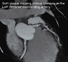
CT can image your coronary and carotid arteries, vessels
to the lower extremities and other organs in addition to
lower extremity veins in patients with suspected deep vein
thrombosis, inside the brain and other parts of the body.
In addition it can image all internal organs.
|
|
These types of images cannot be seen on regular x-ray examinations. CT
makes it possible to diagnose certain diseases earlier and more accurately
than with other imaging tools. Because most diseases are better treated
when found early, appropriate use of CT scans can help save lives.
Computed Tomography is based on the x-ray principal: as x-rays pass through
the body they are absorbed or attenuated (weakened) at differing levels
creating a matrix or profile of x-ray beams of different strength. This
x-ray profile is registered on film, thus creating an image. In the case
of CT, the film is replaced by a banana shaped detector which measures
the x-ray profile. As the x-ray tube and detector make this 360° rotation,
the detector takes numerous snapshots (called profiles) of the attenuated
x-ray beam. Typically, in one 360° lap, about 1,000 profiles are sampled.
Each profile is subdivided spatially (divided into partitions) by the
detectors and fed into about 700 individual channels. Each profile is
then backwards reconstructed (or "back projected") by a dedicated
computer into a two-dimensional image of the "slice" that was
scanned.
Multiple computers are used to control the entire CT system. The main
computer that orchestrates the operation of the entire system is called
the "host computer." There is also a dedicated computer that
reconstructs the "raw CT data" into an image. A workstation
with a mouse, keyboard and other dedicated controls allows the technologist
to control and monitor the exam. The CT gantry and table have multiple
microprocessors that control the rotation of the gantry, movement of the
table (up/down and in/out), tilting of the gantry for angled images, and
other functions such as turning the x-ray beam on an off.
The most modern CT scanner, the one employed at Westside Medical Imaging
has 64-channels increasing the number of snapshots or images obtained
with each cycling of the CT detector. This has finally enabled the CT
scanner to capture diagnostic quality images of the coronary arteries
and other vessels.
CT scanners use x-rays. For your safety, the amount of radiation is kept
to an absolute minimum and our equipment is maintained in top
shape. Because we do not want to expose a developing fetus to
x-ray, be sure to tell your doctor if you are, or think you may
be pregnant before preparing for the CT exam. Please be sure
to let the Westside Medical Imaging receptionist know if there is a likelihood
you may be pregnant. Since some examinations require the use of iodinated
contrast agents, please inform the technician if there you are allergic
to shellfish of if there is any history of contrast or dye allergy or
reaction.
|